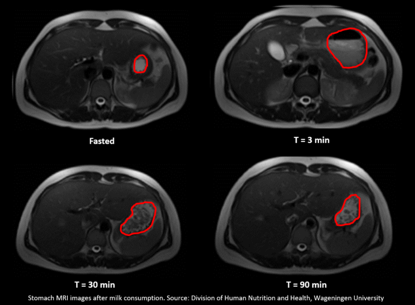
The Paper of the Month for September is from the Proceedings of Nutrition Society and is entitled ‘Monitoring food digestion with magnetic resonance techniques' by Paul Smeets, Ruoxuan Deng, Elise van Eijnatten and Morwarid Mayar.
You have probably heard about MRI, or Magnetic Resonance Imaging, a widely used medical imaging technique that can make 3D images of the entire human body. MRI is based on the same principles as NMR, which stands for Nuclear Magnetic Resonance. NMR is a spectroscopic technique that has been used in chemistry, long before the principles of MRI image formation were developed in the 1970’s, to characterise food composition in detail. Both techniques use a strong magnetic field and precisely tuned radio signals to characterise the object or tissue that is ‘scanned’. MRI stands apart from other medical imaging techniques such as CT because it does not use radiation and is extremely versatile. Ongoing innovation keeps yielding novel MRI techniques that provide benefits such as more detailed images (higher resolution), faster image acquisition or new types of images that can tell us more about the body’s anatomy or function.
At Wageningen University we are studying eating behaviour and digestion from various perspectives. It is not only important what we eat, but also how the foods we ingest are broken down into nutrients that our bodies can be utilised for maintenance and growth. Digestion is a complex series of processes and it is difficult to monitor the fate of foods throughout the digestive tract in detail. Therefore, we and many others use in vitrodigestion models. These allow us to study the breakdown of foods in detail and see how this is affected by e.g. the preparation method or type of ingredient.
In research, MRI has been used for many years to visualise how food ‘behaves’ in the stomach and how fast it moves on into the intestines. This fairly straightforward use of MRI already provides very useful information. We can visualise and often quantify intragastric processes such as layer formation and coagulation (clotting). In our recent review paper in PNS we further argue that magnetic resonance techniques are very well suited to not only study such macroscopic processes in vivo, but that they can also provide more detailed information about the breakdown of nutrients such as proteins. This is not easily achieved, for one thing because MRI, like any other imaging technique, is prone to distortions by movement, and the stomach and intestines are always in motion. However, we have promising data suggesting that by carefully validating MRI markers of digestion in vitro, we can develop novel in vivo MRI approaches that will allow us to literally ‘tune in’ to digestion. This, in turn, can help to refine in vitrodigestion models. Thus, MRI may provide novel tools to strengthen in vitro and in vivodigestion research. This will help us to optimise food properties such as protein digestibility that are important to maximise health benefits.
